By John C. Flucke, DDS
A Decade of 3D Dental Imaging
I’ve been immersed in dental tech for years, and the past decade has been a wild ride with 3D dental imaging reshaping our world. Tools like intraoral scanners and CBCT units have turned complex anatomy into vivid, actionable models, making treatments sharper and patients more engaged than ever.
My First Steps with Digital Impressions
Back in 2006, I got my hands on the original iTero scanner. Picture this: I’m tapping a foot pedal, snapping tooth images one by one, waiting for the software to stitch them into a 3D model. Slow? Sure. Mind-blowing? Absolutely. By 2008, I jumped into CBCT, and suddenly, I could see bone and tissue in ways 2D X-rays never showed. Patients were hooked, asking questions as we explored their scans together, turning consults into real conversations.
Intraoral Scanners: Faster, Smarter, Essential
Today’s intraoral scanners are a different beast. They grab oceans of data in seconds, spitting out live 3D models like magic. Here’s why they’re game-changers:
- Blazing Speed: Full-arch scans in under five minutes.
- Routine Use: New patient scans now happen in hygiene rooms.
- Ease: So intuitive, they’re part of daily flow. I’ve seen offices where these scans are as routine as a cleaning, pulling patients into the digital age with every visit.
CBCT: From Luxury to Must-Have
CBCT has evolved just as fast. Modern units offer:
- Quick Scans: Done in a flash.
- Low Radiation: Safer for everyone.
- AI Smarts: Auto-tracing nerves and segmenting anatomy. Once a shiny toy for early adopters, CBCT is now non-negotiable. Try going back to 2D after seeing a 3D jaw—it’s like ditching color TV for black-and-white.
Innovation to Integration: A Tech Timeline
 Dental tech moves in waves. First, we had “Innovation 1.0”—think clunky carts with intraoral cameras and digital X-rays rolling into operatories. Then came “Integration 1.0” in the late ‘90s, when operatory PCs became the hub, tying tools together. Around 2010, “Innovation 2.0” hit with CBCT and scanners going mainstream. Now, we’re stepping into “Integration 2.0,” where these tools don’t just coexist—they unite.
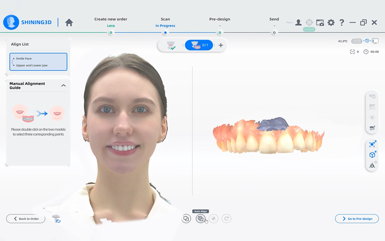
Merging 3D Data: The Virtual Patient Revolution
Imagine this: a CBCT scan, an intraoral scan, and a facial scan, all fused into one 3D model. It’s not sci-fi—it’s here. These virtual patients let me show folks exactly what’s going on inside their mouths, making complex cases feel simple. Virtual smile designs take it further, letting patients preview their new grin right on the screen. Forget old-school wax-ups; this is like showing them a mirror into their future.
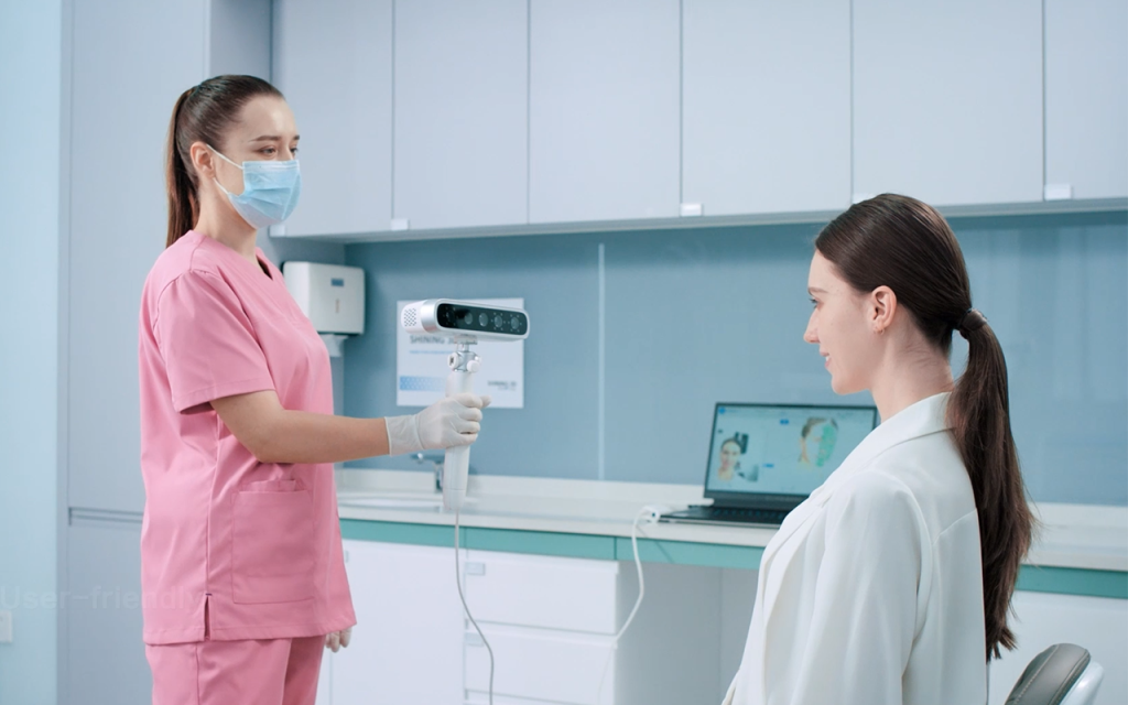
Why MetiSmile Blows My Mind
Shining 3D’s MetiSmile is leading this charge. It uses three cameras, an HD texture lens, and an IR projector to map a patient’s face in moments. Then, it auto-aligns intraoral and CBCT data into a single, rotatable 3D model. Want to geek out? You can tweak transparency to highlight different layers or track jaw movements—centric, open, lateral—for restorations that look and work perfectly. It’s like having a digital crystal ball for treatment planning.
Streamlining Everything
These tools aren’t just cool—they’re practical. They let me:
- Design in-house or send to labs with ease.
- Fabricate restorations with pinpoint accuracy.
- Wow patients with visuals that seal the deal. Consults are smoother, outcomes are better, and everyone’s confidence soars.
What’s Next for Digital Dentistry
Integration 2.0 is just getting started. We’re moving from scattered apps—separate programs for scans, X-rays, charting—to systems that talk to each other. It reminds me of how practice management software tied offices together years ago. This next wave of 3D dental imaging is making dentistry faster, smarter, and more connected, pulling patients into the process like never before. Buckle up—it’s going to be an incredible ride.